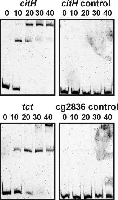
FIG. 8.
Binding of CitB to the promoter regions of citH and tctCBA. DNA fragments (219 to 336 bp; final concentrations, 22 to 35 nM) covering the promoter regions of citH and tctCBA and two DNA fragments serving as negative controls (see the text) were incubated for 30 min at room temperature without CitB (far left lanes) or with a 10- to 40-fold molar excess of purified CitB protein, as indicated, before separation by native polyacrylamide (15%) gel electrophoresis and staining with Sybr green I.

