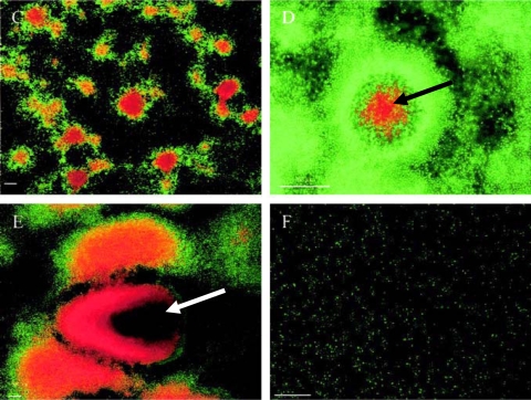
FIG. 14.
Cell death and cell lysis in biofilm dispersal, showing biofilm development and cell death of the P. tunicata wild-type strain. Biofilms were stained with the BacLight Live/Dead bacterial viability kit. Red propidium iodide-stained cells have a compromised cell membrane and are dead. Time points after inoculation are shown as follows: (C) 48 h; (D) 72 h; (E) 144 h; (F) 168 h. Cell death can be observed at 48 h, and cell lysis (arrow in panel D) and extensive cell death (arrow in panel E) are seen at 144 h, prior to complete dispersal of the biofilm at 168 h. Bars, 50 μm. (Reprinted from reference 210 with permission.)

