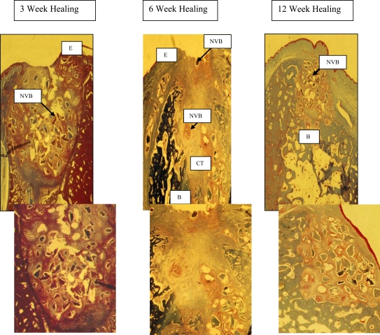
Fig. (2). DFDBA and Membrane.
3 weeks - the specimens have a thin epithelium (E) overlying the extraction site with a slight discontinuity. The socket was still filled with non-vital DFDBA particles (NVB) and dense inflammatory infiltration. No new bone formation was seen in the extraction socket.
6 weeks - epithelialization (E) of the wound was not complete and non-vital osseous particles appeared to be present in the fibrous connective tissue overlying the area. The socket contained fibrous connective tissue (CT) but non-vital osseous particles surrounded by an inflammatory infiltrate could still be noted. There was a minimal amount of new bone (B) observed in the apical third of the socket.
12 weeks - maxillary sites had complete fill of the socket with new bone and only a few bone chips were seen in the overlying connective tissue. In mandibular sites (above section), new bone (B) filled most of the socket, but in the coronal third there was a cone shaped area of non-vital bone particles (NVB), surrounded by inflammatory infiltrate and connective tissue. This cone was narrow in the center of the socket becoming wider in the coronal portion of the area.

