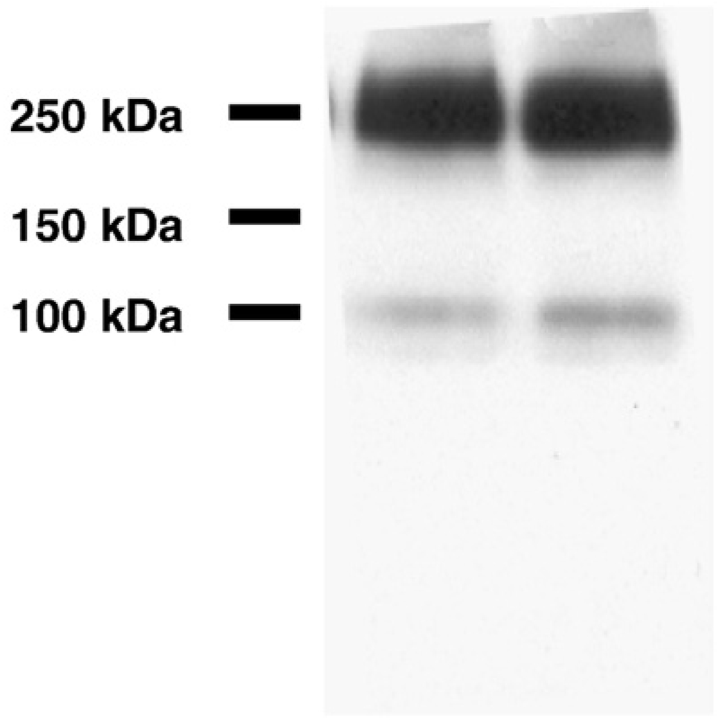
Fig. 1.
The expression of mGluR2 in rat primary cortical neurons (DIV 7–10). The membrane fraction rat primary cortical neurons were separatedon a 7.5% acrylamide gel and mGluR2 was detected by anti-mGluR2 rabbit polyclonal antibody. As expected, the monomer (~100 kDa) and dimer (~200 kDa) are observed and this staining pattern is the same as found for mGluR2 in human brain samples.

