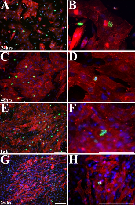
Fig. 4.
Immunofluorescent images of embryonic myocytes co-labeled for phospho-histone H3 (green, mitosis marker) and α-sarcomeric actinin (red, myocyte marker). Phospho-histone H3 cells are seen for up to 2 weeks after isolation. Nuclei (blue) are labeled with bis-benzimide. Images were taken at 24 h (a, b), 48 h (c, d), 1 week (e, f), and 2 weeks (g, h) after isolation. Scale bars = 100 μm

