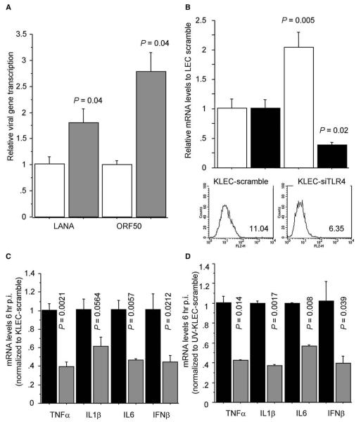
Figure 2. Increased Susceptibility of Cells Lacking TLR4 to KSHV Infection.
(A) mRNA levels of KSHV-encoded LANA-1 and ORF50 transcripts after KSHV infection (48 hr p.i.) of peritoneal macrophages from C57BL10/ScSnJ mice (wild-type, white bars) and C57BL10/ScNJ mice (Tlr4−/−, gray bars). Levels normalized to average gene expression in KSHV-infected C57BL10/ScSnJ macrophages. P values indicate statistical significance of changes between C57BL10/ScNJ and C57BL10/ScSnJ macrophages.
(B) LANA-1 mRNA levels (white bars) in KLEC (48 hr p.i.). LEC were transfected with nontargeting siRNA (KLEC scramble) or TLR4-targeting siRNA (KLEC-siTLR4) 48 hr before KSHV infection. TLR4 surface expression (histograms; TLR4 geometrical mean fluorescence shown) and mRNA levels (black bars) were determined 48 hr post siRNA transfection. mRNA levels are normalized to KLEC-scramble levels.
(C) TNF-α, IL1-β, IL-6, and IFN-β mRNA levels 6 hr after KSHV infection of KLEC scramble (black bars) or KLEC-siTLR4 (gray bars). Levels are normalized to KLEC scramble.
(D) As in (C), but after exposure of LEC to UV-inactivated KSHV. In (B)–(D), P values indicate statistical significance of changes between mRNA levels in KLEC scramble and KLEC-siTLR4. In all panels, error bars represent standard deviation.

