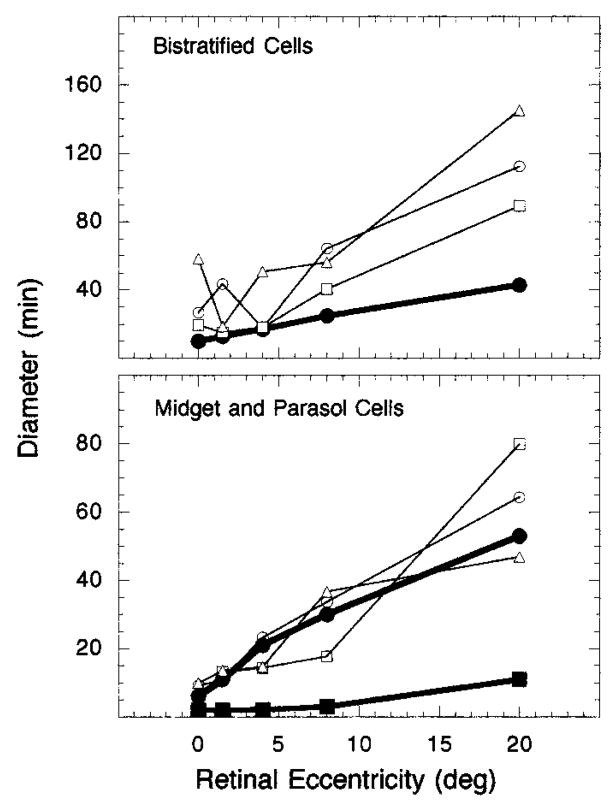
Fig. 9.
Diameter of Ricco’s area at the retina and dendritic field size plotted as a function of retinal eccentricity: Top, diameter of Ricco’s areas (open symbols) for an S-cone mechanism compared with dendritic field size of B/Y bistratified ganglion cells (filled circles); bottom, diameter of Ricco’s areas (open symbols) for an L-cone mechanism compared with dendritic field sizes of midget (filled squares) and parasol (filled circles) ganglion cells. Each set of open symbols represents data from a different observer.

