Figure 2.

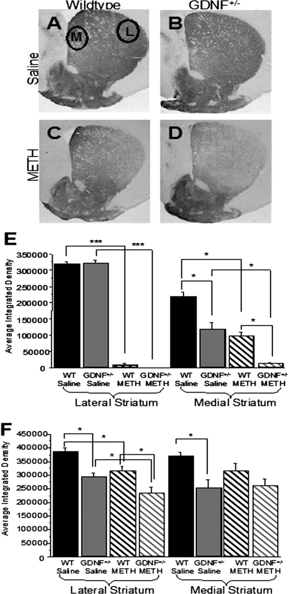
After METH treatment, 3- and 12-month-old GDNF+/− mice demonstrated a greater loss of striatal TH-IR than WT mice. A–D, Photomicrographs of TH-IR in coronal hemisections of the striatum (medial to the left; lateral to the right) from 3-month-old saline-treated WT mice (A), saline-treated GDNF+/− mice (B), METH-treated WT mice (C), and METH-treated GDNF+/− mice (D). E, Quantitation of the average integrated density from the four treatment groups at 3 months of age confirmed that there were no differences in the lateral region (right) of the striatum between the GDNF+/− and WT mice treated with saline, and METH induced a similar TH-IR depletion in the lateral striatum of GDNF+/− and WT mice when compared with saline-treated mice (***p < 0.001). In the medial striatum (left), GDNF+/− mice expressed less TH-IR than WT mice treated with saline (*p < 0.05), and METH induced a greater depletion in the medial striatum of GDNF+/− than WT mice (*p < 0.05). F, Quantitation of the average integrated density of TH-immunoreactive sections from the four treatment groups at 12 months of age demonstrated that TH-IR levels were significantly less in both the lateral and medial striatum of GDNF+/− mice versus WT mice treated with saline (*p < 0.05), that there was a residual deficit in TH-IR in METH- versus saline-treated WT mice in the lateral region of the striatum (right; *p < 0.05), and that there was significantly less TH-IR in the lateral striatum of GDNF+/− versus WT mice treated with METH (*p = 0.05). Circles denote measurement areas of the lateral (L) and medial (M) regions of the striatum. n = 8 per group. Error bars indicate SEM.
