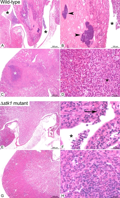
FIG. 8.
Histopathological analyses of kidneys in mice infected with either S. aureus wild-type strain SH1000 (A to D) or its isogenic Δstk1 mutant (E to H). (A) Low magnification of the renal crest (white star) and pelvis (black star), with numerous infiltrates of neutrophils destroying the normal tubular architecture of the tissue. (B) Higher magnification of the renal crest showing bacterial colonies (arrowheads) in a necrotic area (ischemic necrosis). A neutrophilic infiltrate is also observed in the pelvis (black star). (C) Focal abscess in the renal cortex (white star). (D) Higher magnification of the abscess organization, with centrally located fragmented (i.e., karyorrhectic) neutrophils and cell debris (suppuration) (white star) and a peripheral rim of numerous macrophages associated with lymphocytes and plasma cells (black star). (E) Low magnification of the renal crest and pelvis showing no significant histological lesions. (F) Higher magnification of the renal papilli showing a small neutrophilic infiltrate in the pelvis (black star) extending to the overlying epithelium (arrow). (G) Low magnification of the renal cortex with no significant histological lesions. (H) In one mouse, small (less than 150 μm in diameter) infiltrates of macrophages associated with less numerous lymphocytes and plasma cells were observed in the cortex.

