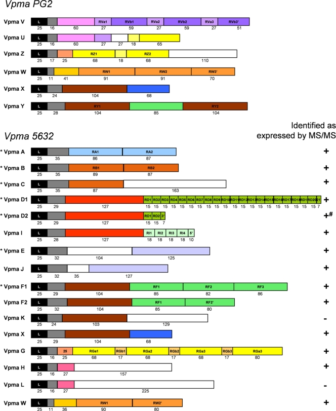
FIG. 2.
Structural features and comparison of vpma gene products in M. agalactiae strains PG2 and 5632. Predicted Vpma proteins are represented schematically by boxes and begin with a homologous 25-amino-acid leader sequence (black boxes) followed by regions that have homology between vpma gene products or that are repeated within the same product (colored boxes). Two boxes of the same color display an amino acid identity of >30%. White boxes represent unique sequences. Numbers below boxes indicate the numbers of amino acids. For strain 5632, detection by MS/MS of expressed specific Vpma peptides is noted (+). −, no specific peptides were detected for the corresponding Vpma; +#, VpmaD2 peptides detected are not specific because all are shared with VpmaD1 or -I (see the list of detected peptides in Table S3 in the supplemental material). An asterisk indicates that the corresponding vpma gene is present in 5632 at both vpma loci.

