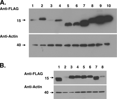
FIG. 2.
Western blot analysis of L mutant and chimeric proteins. (A) Western blot analysis of DA and GDVII L mutant proteins. All of the constructs shown in Fig. 1B were subcloned into p3xFLAG-CMV14 to be expressed as FLAG-tagged fusion proteins in BHK-21 cells. The transfected cells were harvested at 20 h after transfection, and total cell lysates were prepared in the lysis buffer. The cell lysates (10 μg) were immunoblotted with anti-FLAG antibody. Lanes 1, 3, 5, 7, and 9 represent Lwt, LΔZ, LΔA, LΔS/T, and LΔC, respectively, of the DA strain. Lanes 2, 4, 6, 8, and 10 represent Lwt, LΔZ, LΔA, LΔS/T, and LΔC, respectively, of the GDVII strain. Immunoblotting for actin using the same cell lysates also is shown as a loading control. (B) Western blot analysis of DA and GDVII L chimeric proteins. The chimeric mutants shown in Fig. 1C and D were subcloned into p3xFLAG-CMV14 or p3xFLAG-CMV10, and protein expression was analyzed by Western blotting as described for panel A. Lanes 1 and 2 represent Lwt of DA, and lanes 3 and 4 represent Lwt of GDVII. Lanes 5 and 6 represent chimera D/G, and lanes 7 and 8 represent chimera G/D. Lanes 1, 3, 5, and 7 represent the proteins expressed as FLAG-tagged proteins at the C terminus, and lanes 2, 4, 6, and 8 represent the proteins expressed as FLAG-tagged proteins at the N terminus. Immunoblotting for actin using the same cell lysates also is shown as a loading control.

