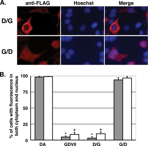
FIG. 6.
Subcellular localization of chimeric L proteins of DA and GDVII strains. (A) Subcellular localization of chimera D/G and G/D. BHK-21 cells were transfected with the expression plasmids for FLAG-tagged chimera D/G or G/D at the C terminus. The cells were fixed 20 h after transfection and stained with anti-FLAG antibody for L and its mutants (anti-FLAG). Antibody-antigen complexes were detected with Alexa Fluor 594-conjugated anti-mouse IgG. Nuclei were stained with Hoechst 33258 (Hoechst). Cells were observed by a fluorescence microscope (Axiovision; Carl Zeiss). Merge represents merged images stained with anti-FLAG and Hoechst. (B) Distributions of the chimeras D/G and G/D. Histograms show the percentages of the transfected cells showing both nuclear and cytoplasmic FLAG staining. None of the cells showed solely nuclear staining. The localization was observed by fluorescence microscopy in five fields containing 10 to 50 cells positive for FLAG expression. Closed and open columns showed the results for the proteins FLAG tagged at the C and N termini, respectively. Each value represents the means ± standard errors of the means from three independent experiments. *, significant between DA and GDVII or D/G tagged at the C terminus at P < 0.0001; +, significant between DA and GDVII or D/G tagged at the N terminus at P < 0.0001.

