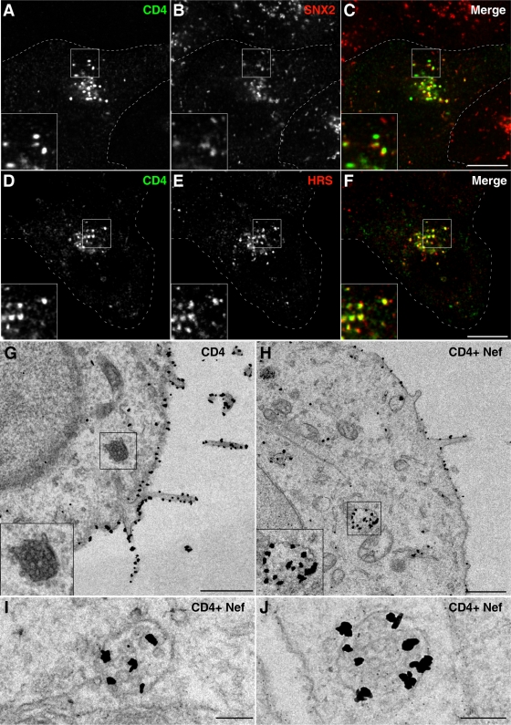
FIG. 2.
Nef induces redistribution of CD4 to multivesicular bodies. (A to F) HeLa cells transfected with pCMV-CD4 and pCIneo-Nef-wt were fixed 16 h later and costained with mouse monoclonal antibody to CD4 and rabbit polyclonal antibodies to either SNX2 (A to C) or HRS (D to F), followed by Alexa-488-conjugated donkey antibody to mouse IgG (green channel) and Alexa-594-conjugated donkey antibody to rabbit IgG (red channel). Cells were imaged by confocal laser scanning microscopy. Cell outlines are indicated by dashed lines. Yellow in the merged images indicates colocalization. Bar, 10 μm. The insets represent the boxed areas at a magnification of ×2. (G to J) HeLa cells were transfected with pCMV-CD4 (G) or pCMV-CD4 plus pCIneo-Nef-wt (H to J). After 16 h, cells were fixed and processed for immunoelectron microscopy as described in the Materials and Methods section. Examples of MVBs are shown in the insets of panels G and H and in panels I and J. The insets in panels G and H show the boxed areas at a magnification of×2. Bars, 1 μm (G and H) 0.2 μm (I and J).

