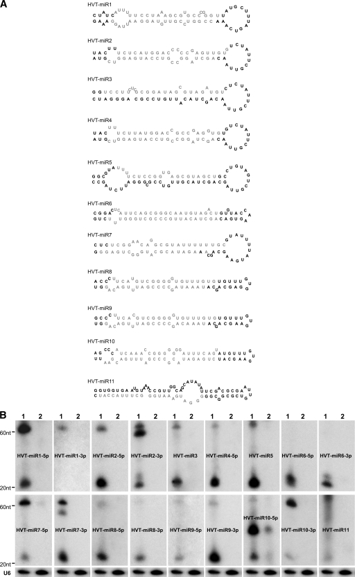
FIG. 2.
Identification of cloned HVT miRNAs. (A) Secondary structures of HVT pre-miRNAs predicted using the MFOLD algorithm. The mature miRNA strands are indicated in light gray. (B) Northern blot analysis demonstrating the expression of HVT miRNAs. Total RNAs from HVT-infected CEFs (lanes 1) and uninfected CEFs (lanes 2) were separated on a 15% denaturing polyacrylamide gel and probed with [γ-32P]ATP-radiolabeled antisense oligonucleotides to the indicated miRNAs. Size markers indicate the positions of the pre-miRNA and the mature miRNA. The cellular U6 small nuclear RNA served as the loading control. A representative blot of this set is shown.

