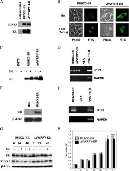
FIG. 5.
RUNX3-ER and ΔVWRPY-ER both translocate to the nucleus and bind to the RUNX1 P1 promoter. (A) HEK293 cells were transfected with plasmid pCEP4-RUNX3-ER or pCEP4-ΔVWRPY-ER, and cells were lysed in sample buffer and fractionated by SDS-PAGE. Western blotting was performed, probing for RUNX3 or ER. (B) HEK293 cells were transfected with the pCEP4-RUNX3-ER or pCEP4-ΔVWRPY-ER expression plasmid, and after 24 h, 5 μM β-estradiol (Est) was added. Twenty-four hours after β-estradiol addition, cells were fixed and probed with an anti-ER antibody followed by fluorescein isothiocyanate (FITC)-conjugated anti-rabbit secondary antibody. The locations of the fusion proteins were assessed by confocal microscopy. (C) DG75 cells stably transfected with pCEP4-RUNX3-ER or pCEP4-ΔVWRPY-ER were incubated with and without 5 μM β-estradiol for 24 h. RIPA extracts were prepared, and 50 μg protein was analyzed by Western blotting, probing for the ER component of the RUNX-ER fusion protein. (D) DG75 pCEP4-RUNX3-ER or pCEP4-ΔVWRPY-ER stable cells were incubated with 5 μM β-estradiol for 24 h. A ChIP assay was performed on extracts from these cells by using the ER antibody, a nonspecific negative-control antibody (NSA), and an RNA polymerase II (Pol II) positive control. PCR amplification using primers for the RUNX1 P1 promoter (R1P1) or the GAPDH promoter was performed on the immunoprecipitations, and products were fractionated by agarose gel electrophoresis. (E) Extracts of IB4 cells or IB4 cells stably expressing RUNX3-ER (as in Fig. 2), treated with estrogen, were analyzed by Western blotting with an ER antibody for RUNX3-ER expression. (F) The estrogen-treated IB4 cells expressing RUNX3-ER were tested in the ChIP assay, using either an ER antibody (RUNX3-ER), a nonspecific control antibody (NSA), or an RNA Pol II antibody. PCR was for the RUNX1 P1 promoter or the GAPDH promoter. (G) DG75 pCEP4-RUNX3-ER or pCEP4-ΔVWRPY-ER cells were incubated with 5 μM β-estradiol, RIPA extracts were prepared, and 50 μg protein was separated by SDS-PAGE. Western blotting was performed, probing for ER, RUNX1, and β-actin. (H) DG75 pCEP4-RUNX3-ER or pCEP4-ΔVWRPY-ER cells were incubated with 5 μM β-estradiol, harvested, and counted at the indicated times. Results are expressed as average total numbers of cells in a 10-cm dish.

