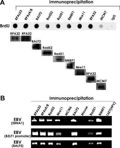
FIG. 5.
HRR factors such as phosphorylated RPA32, Mre11, Rad 51, and Rad52 are loaded onto EBV genomes. (A) Association of newly synthesized DNA with RPA32 Ser-33, RPA32 Ser-4/Ser-8, Rad52, Rad51, BMRF1, Mre11, and RPA32. Lytic replication was induced in Tet-BZLF1/B95-8 cells with 2 μg/ml of doxycycline, and newly synthesized DNAs were labeled for 1 h with 15 μM BrdUrd at 24 h p.i. Nuclear extracts were prepared as described in Materials and Methods and subjected to IP with each of the anti-RPA32 Ser-33, anti-RPA32 Ser-4/Ser-8, anti-Rad52, anti-Rad51, anti-BMRF1, anti-Mre11, and anti-RPA32 antibodies. An anti-MCM7 antibody and nonimmune mouse IgG were also applied as negative controls. The proteins and DNAs were eluted from the beads using SDS and EDTA, and DNAs were then purified with Chroma Spin-10 gel filtration columns (Clontech Laboratories, Inc.) and dot blotted onto Hybond N+ membranes. The BrdUrd (BrdU)-incorporating DNA in the precipitate was immunostained with an anti-BrdUrd antibody, and the included proteins were subjected to immunoblot analysis with the specific antibodies indicated at the top. (B) IP of the EBV genome by anti-RPA32 Ser-33, anti-RPA32 Ser-4/Ser-8, anti-Rad52, anti-Mre11, anti-BALF2, anti-Rad51, and anti-BMRF1 antibodies. Tet-BZLF1/B95-8 cells were cultured in the presence of 2 μg of doxycycline/ml and harvested at 24 h p.i. Nuclear extracts were prepared as described in Materials and Methods and subjected to IP using anti-RPA32 Ser-33, anti-RPA32 Ser-4/Ser-8, anti-Rad52, anti-Rad51, anti-BMRF1, anti-Mre11, and anti-BALF2 protein-specific antibodies, respectively. Nonimmune mouse IgG and anti-MCM6 or anti-16E7 antibodies were applied as negative controls. The proteins and DNAs were eluted from the beads using SDS and EDTA, and DNAs were then purified through Chroma Spin-10 gel filtration columns and amplified by PCR using primers specific for the EBV EBNA1 gene, the BZLF1 promoter, and the BALF5 gene. The amount of sample used in the PCR of the immunoprecipitated lane represents 1.2% of the precipitated DNA. These experiments were repeated three times, and the PCRs were performed in triplicate.

