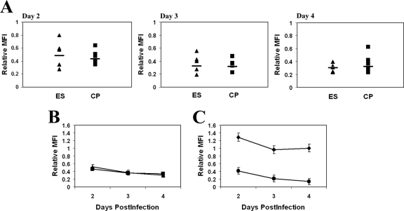
FIG. 2.
(A) Relative MFIs of HLA-A*02 on cells infected with isolates from five ES (triangles) and eight CP (squares) on days 2 to 4 postinfection. The relative MFI is defined as the MFI of the infected cells divided by the MFI of the uninfected CD4+ T cells in each sample. The horizontal bars represent the median for each group. (B) Average relative MFI of HLA-A*02 for cells infected with isolates from ES and CP on each day. (C) Average relative MFI of HLA-A*02 for cells infected with the wild-type NL4-3-green fluorescent protein virus (diamonds) or the Nef− Vpr− mutant virus (circles).

