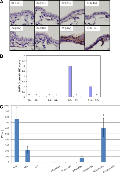
FIG. 4.
IHC detection of HMPV infection and lung virus titers following glucocorticoid reactivation of HMPV. Lung sections were collected from mice at the times indicated after Dx (+Dx) or PBS (−Dx) treatment and stained with rabbit antisera reactive to HMPV G protein. (A) Images (×100 magnification) of the lung sections are shown. (B) To aid interpretation, the IHC scores are indicated as follows: 0, no IHC staining detected or negative; 0.5, <10% cells have detectable IHC staining; 1, weak positive where 10 to 20% cells have detectable IHC staining; 2, positive where 30 to 40% cells have detectable IHC staining; 3, positive where 50 to 70% cells have detectable IHC staining; and 4, strong positive where 80 to 100% cells have detectable IHC staining. +, Dx treatment; −, no Dx treatment. (C) Lung virus titers steroid activation. Lung virus titers were determined at days 4, 7, or 13 posttreatment by using immunostaining plaque assay as previously described (1). The error bars indicate the standard error of the mean. The asterisk indicates significant (P < 0.05) differences between day 13 and day 4 lung virus titers after Dx treatment.

