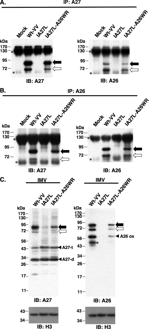
FIG. 2.
Immunoprecipitation (IP) of A26-A27 protein complexes from cell lysates. BSC40 cells were infected with viruses as shown at the top at an MOI of 10 PFU/cell and were harvested at 24 h p.i. Cell lysates were immunoprecipitated with anti-A27 Ab (1:100) (A) or anti-A26 Ab (1:50) (B). The immunoprecipitates were washed, boiled, and separated on SDS-PAGE and analyzed by immunoblotting using anti-A26 and anti-A27 Abs as described for panel A. The black and white arrows indicate the 90- and 70-kDa protein complexes, while small asterisks indicate Ab heavy and light chains. (C) A26-A27 protein complexes were stably incorporated in IMV particles. Equivalent amounts of each IMV were separated by SDS-PAGE and analyzed by immunoblotting with anti-A26 (1:1,000), anti-A27 (1:1,000), and anti-H3 (1:1,000) Abs. The black and white arrows indicate the 90- and 70-kDa protein complexes, respectively. The asterisk represents the degraded form of A26 protein. IB, immunoblot.

