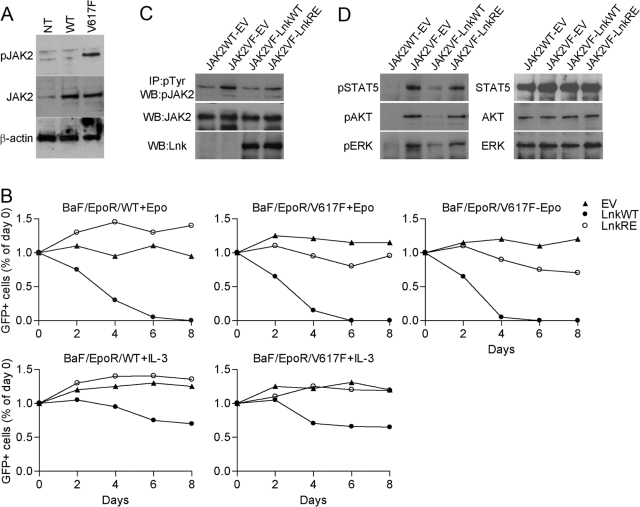
Fig. 1.
Lnk inhibits proliferation of hematopoietic cells expressing JAK2V617F. (A) Western blot analysis of JAK2 total protein and phosphorylation levels in the Ba/F3-EpoR cells stably expressing JAK2WT (BaF/EpoR/WT) or JAK2V617F (BaF/EpoR/V617F). Cells were depleted of cytokines for 16 h. NT, Nontransfected Ba/F3-EpoR cells. (B) BaF/EpoR/WT and BaF/EpoR/V617F cells were transduced with MSCV-IRES-GFP (MIG) empty vector (EV), MIG WT Lnk (LnkWT), or MIG SH2 mutant Lnk (LnkRE). Two days later (Day 0), the percent of transfected cells was analyzed by measuring GFP (considered as 100%). Subsequently, GFP expression was measured and calculated relative to that of Day 0. Epo (10 U/ml) and IL-3 (10 ng/ml) were added as indicted. Results are representative of three independent experiments. (C and D) BaF/EpoR/WT and BaF/EpoR/V617F cells were transduced with the MIG vectors and cultured for 2 days with (BaF/EpoR/WT) or without Epo (BaF/EpoR/V617F), after which, both cell lines were incubated without Epo for 16 h. FACS-sorted GFP-positive cells were lysed. (C) Levels of pJAK2 were analyzed by immunoprecipitation (IP) with pTyr antibody, followed by Western blot (WB) with pJAK2 antibody. Levels of total JAK2 and Lnk were analyzed by Western blot. (D) Western blot analysis of phosphorylation and total levels of the indicated proteins. Experiments were repeated three times with similar results.

