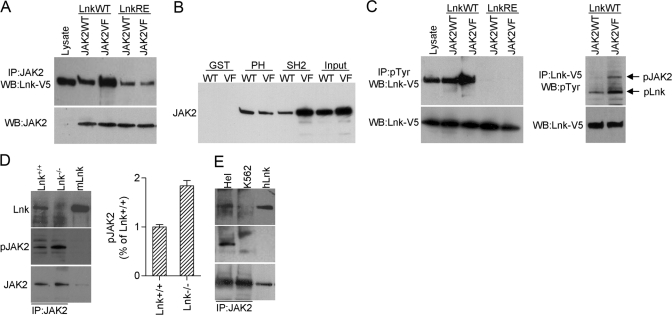
Fig. 2.
Lnk interacts with JAK2WT and JAK2V617F. (A–C) 293T cells were cotransfected with combinations of JAKWT (JAK2WT), JAK2V617F (JAK2VF), WT Lnk-V5 (LnkWT), or SH2 mutant Lnk-V5 (LnkRE) as indicated. Lysates were immunoprecipitated with JAK2, pTyr, or V5 antibodies and analyzed by Western blot as indicated (A and C, upper panels). JAK2 and Lnk levels in the lysates were analyzed by Western blot with JAK2 (A) and V5 (C) antibodies (lower panels). Lysate from 293T-Lnk-V5-transfected cells was used as control for molecular size (A and C, lysate, first lane). (B) Lysates from 293T cells transfected with JAK2WT or JAK2V617F were incubated with GST, GST-Lnk PH (PH), or GST-Lnk-SH2 (SH2) fusion proteins. GST-protein complexes were analyzed by Western blot with JAK2 antibody. Input represents one-tenth of the lysate used for the pull-downs. (D) BM cells from Lnk+/+ and Lnk−/− mice were starved in RPMI 1640 for 4 h and stimulated with 20 ng/ml SCF, 10 U/ml Epo, and 20 ng/ml Tpo for 30 min. Lysates were immunoprecipitated with JAK2 antibody. Bar graph shows the mean ± sd of pJAK2 from two experiments. Results are expressed as a relative percentage compared with pJAK2 in Lnk+/+ cells. (E) Lysates from Hel and K562 cells were immunoprecipitated with JAK2 antibody. (E and D) Blots were probed with the indicated antibodies. Lysates from 293T Lnk-transfected cells were used to control for molecular size. mLnk, Murine Lnk; hLnk, human Lnk. Shown are representative results from two independent experiments.

