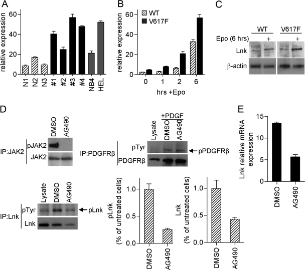
Fig. 3.
High levels of Lnk are associated with MPD. (A) Lnk expression was measured by real-time PCR in CD34+ hematopoietic progenitor cells from MPD patients (JAK2V617F-negative #1 and #2; JAK2V617F-positive #3 and #4), as well as normal donors (N1–N3) and cell lines NB4 (JAK2WT) and Hel (JAK2V617F). Relative Lnk mRNA levels are expressed in arbitrary units as a ratio of Lnk transcripts:18S transcripts. Data represent the mean ± sd of triplicate samples. (B and C) Real-time PCR (B) and Western blot (C) analysis of Lnk expression in Ba/F3 cells stably expressing EpoR and JAK2WT (WT) or JAK2V617F (V617F). Cells were depleted of cytokines for 16 h and then stimulated with Epo (10 U/ml) for the indicated times. Experiments were repeated three times. (D and E) Hel cells were treated for 4 h with JAK2 inhibitor (AG490, 50 μM) or vehicle alone (DMSO). (D) pJAK2 was analyzed by immunoprecipitation with JAK2 antibody followed by Western blot with pJAK2 antibody; JAK2 protein level was analyzed by Western blot. pLnk was analyzed by immunoprecipitation with pTyr antibody followed by Western blot with Lnk antibody; total Lnk levels were analyzed by Western blot. Bar graphs show the mean ± sd of pLnk and Lnk from three experiments. Results are expressed as a relative percentage compared with untreated cells. As a negative control, pPDGFRβ was analyzed in cells stimulated with PDGF-BB (20 ng/ml) for the last 5 min of AG490/DMSO treatment. Lysates from 293T Lnk- and PDGFRβ-transfected cells were used to control for molecular size. (E) Lnk mRNA levels were measured by real-time PCR. Relative Lnk mRNA levels are expressed in arbitrary units as a ratio of Lnk transcripts:18S transcripts. Data represent the mean ± sd of triplicate samples. Shown are representative results from three experiments.

