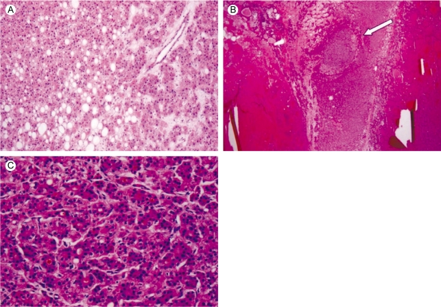
Figure 4.
Microscopic findings. (A) The liver cell cords are one to two cells thick and the cell density is slightly increased compared to the surrounding liver. Portal tracts are absent and focal steatosis is noted (haematoxylin-eosin stain, ×100). (B) A low power microscopic view reveals hepatocellular carcinoma (white arrow) arising in the hepatocellular adenoma (haematoxylin-eosin stain, ×12.5). (C) Microscopic findings of the hepatocelllular carcinoma area. Note the typical trabecular features are more than three cell in thickness (Edmondson-Steiner grade II), and pseudoacinar formation is also noted (haematoxylin-eosin stain, ×200).

