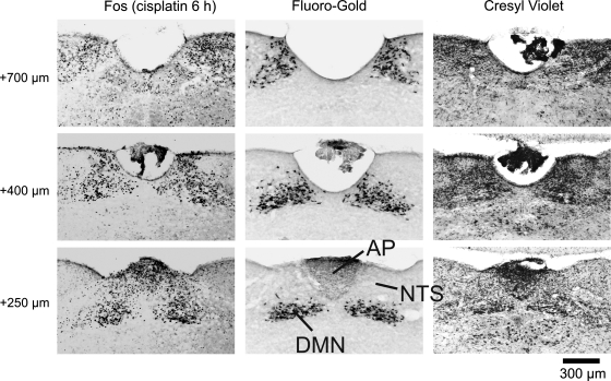
Fig. 1.
Representative images from the Suncus murinus hindbrain showing the rostral extent of Fos expression induced by cisplatin treatment in the dorsal vagal complex (DVC). Areas analyzed for Fos expression included the nucleus of the solitary tract (NTS), area postrema (AP), and dorsal motor nucleus (DMN). Six hours after cisplatin (30 mg/kg ip) treatment, Fos expression (left) was increased at several levels (+250, +400, and +700 μm) rostral to the obex. The boundaries of hindbrain areas were determined by retrograde labeling of the DMN with Fluoro-Gold (center) and cresyl violet staining of cytoarchitecture (right).

