Figure 3.
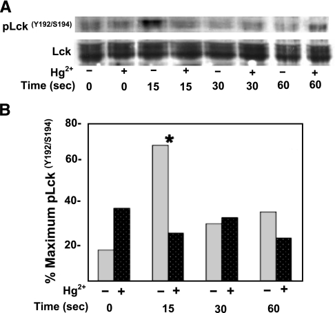
Hg2+ inhibits Lck(Y192/S194) phosphorylation after TCR stimulation. A) Jurkat T cells were incubated in RPMI with (+) or without (−) μM HgCl2, followed by the addition of α-CD3 antibody to activate TCRs. Aliquots containing equivalent cell numbers were incubated at 37°C, and reactions were stopped at the times indicated. Cells were then lysed in SDS, and phosphorylated Lck was detected by Western blotting with an antibody specific to Lck dually phosphorylated on tyrosine 192 and serine 194. The blots were then stripped and probed for total Lck. Results are representative of 7 independent experiments. B) A quantitative analysis of pLck(Y192/S194) after TCR signaling in cells that had been treated with mercury (dark gray, dotted bars) and those that had not (light gray bars) (performed as described for Fig. 2) was undertaken. Results from 7 independent experiments were averaged. Results between Hg2+-treated and untreated cells are only statistically different for the 15-s time point. *P < 0.05; paired Student’s t test.

