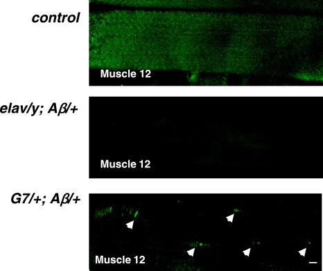
Figure 7.
Endogenous Aβ42 fibrils revealed by thioflavin-S staining in muscle fibers of G7/+; Aβ/+ larvae. Thioflavin-S-positive staining was detected in the muscle cells in G7/+; Aβ/+ larvae, but not in the ctrl and elav/y; Aβ/+ larvae. Arrowheads indicate fiber-like staining within muscle cells. Scale bar = 10 μm.

