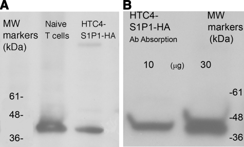
Figure 5.
Western blots of SDS-polyacrylamide gradient gel electrophoretic patterns. A) Proteins extracted from normal human T cells (left lane) and S1P1-HA-transfected HTC4 cells (right lane) were electrophoresed, blotted, and labeled with patient MAW plasma Igs followed by mouse anti-human IgG (H+L) and then HRP-conjugated donkey F(ab′)2 anti-mouse IgG (H+L). B) Proteins extracted from S1P1-HA-transfected HTC4 cells were preabsorbed on Sepharose-rat monoclonal anti-HA Ab and eluted, and then 10 and 30 μg of absorbed proteins was electrophoresed, blotted, and labeled with patient MAW plasma Igs, followed by mouse anti-human IgG (H+L) and then HRP-conjugated donkey F(ab′)2 anti-mouse IgG (H+L).

