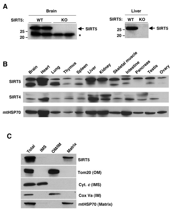
Figure 1. SIRT5 is broadly expressed and localized in the mitochondrial matrix.
(A) The SIRT5 antibody specificity was tested by western blotting of mouse total brain (left) and mouse liver mitochondria matrix (right) lysates from SIRT5 wild type and KO mice. Arrow indicates SIRT5 and asterisk indicates non-specific band.
(B) Total tissue lysates were subject to western blotting using anti-SIRT5, anti-SIRT4 and anti-mtHSP70 antibodies.
(C) Mitochondria were isolated from mice liver and subject to sub-mitochondrial fractionation. Blots are probed with antibodies to SIRT5, Tom20 - an outer membrane (OM) marker, Cytochrome c - an inter membrane space (IMS) marker, COX Va - an inner membrane (IM) marker and mtHSP70, a matrix marker.

