FIG. 2.
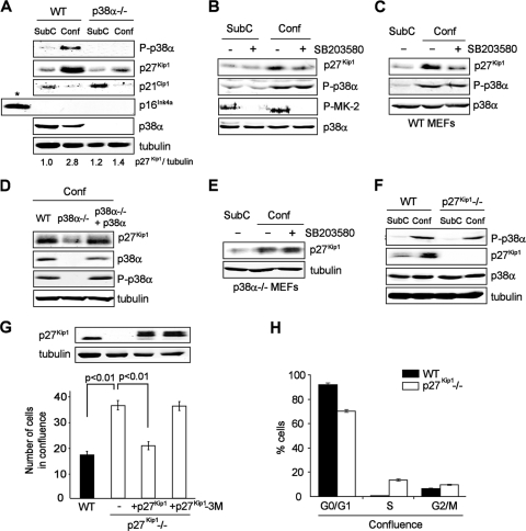
p27Kip1 mediates the antiproliferative effect of p38α in contact inhibition. (A) Immunoblot analysis of subconfluent (SubC) and confluent (Conf) WT and p38α−/− MEFs. Primary MEFs were used as a control for p16Ink4a expression (indicated by an asterisk). p27Kip1 protein levels were quantified by densitometry and normalized to tubulin. (B) Mouse NIH 3T3 fibroblasts were grown to subconfluence (SubC) or confluence (Conf), treated overnight with the p38α and p38β inhibitor SB203580 where indicated (+, present; −, absent), and analyzed by immunoblotting. P-MK-2, phospho-MAPK-activated protein kinase 2. (C to E) WT and p38α−/− MEFs and p38α−/− MEFs with p38α added back were grown to subconfluence (SubC) or confluence (Conf), treated overnight with SB203580 as indicated (+, present; −, absent), and analyzed by immunoblotting. (F) Sparse (SubC) and confluent (Conf) WT and p27Kip1−/− MEFs were analyzed by immunoblotting. (G) WT and p27Kip1−/− MEFs stably transduced with WT p27Kip1, mutant p27Kip1-3M, or empty vector (−) were grown to confluence and counted. Total lysates were analyzed by immunoblotting. Values are numbers of cells in 10,000s. (H) Percentages of confluent WT and p27Kip1−/− MEFs in the different phases of the cell cycle were determined by flow cytometry. Error bars indicate standard deviations of results for biological replicates. P values were determined by Student's t test and indicate 99% error-free (P < 0.01) statistical significance.

