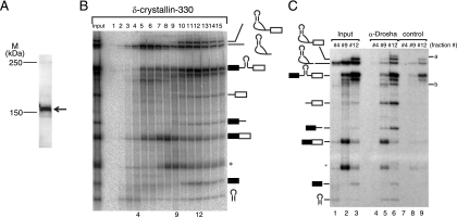
FIG. 6.
Identification of Drosha-associated RNAs. (A) Western blotting of HeLa cell nuclear extracts with anti-Drosha antibody. The band corresponding to Drosha is indicated by an arrow. The positions of size markers are indicated on the left. (B) Fractionation of in vitro splicing mixture by glycerol gradient centrifugation. In vitro splicing was performed with δ-crystallin-330 pre-mRNA and a 20-min incubation. The input lane contained 5% of the total splicing mixture. The structure of each RNA product is shown schematically at the right side of the panel. (C) Immunoprecipitation of RNAs from the fractions shown in panel B. The identities of RNA products are shown on the left side of the panel. The unidentified RNAs precipitated efficiently by anti-Drosha antibody are indicated on the right (a and b). The input lanes contained 5% of the fractions.

