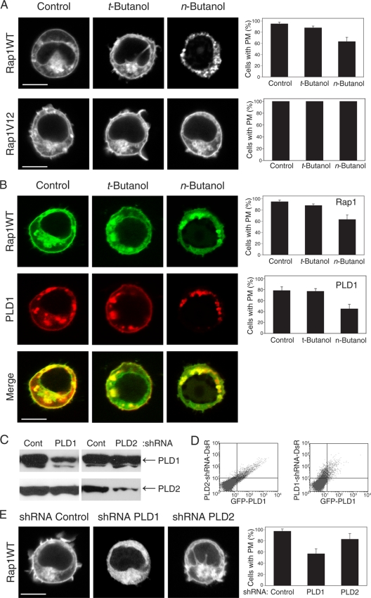
FIG. 2.
Rap1 expression on the plasma membrane depends on PLD1. (A) Inhibition of PLDs with n-butanol affects the plasma membrane (PM) localization of GFP-Rap1 but not GFP-Rap1V12. (B) n-butanol inhibits anti-CD3-stimulated translocation of both GFP-Rap1 and RFP-PLD1 from cytoplasmic vesicles to the plasma membrane. (C) Endogenous PLD1 and PLD2 in HeLa cells, detected by immunoblotting before and 72 h after expression of the indicated shRNA. (D) PLD1 knockdown in Jurkat cells, shown by cytofluorimetry of cells expressing GFP-PLD1 (x axis) and the indicated shRNA, which also directs expression of RFP from an internal ribosome entry site (y axis). (E) Silencing PLD1 but not PLD2 expression with shRNA inhibits plasma membrane localization of GFP-Rap1. Images show representative cells (bars, 5 μm), and the bar graph shows means ± standard errors of the means (SEM) (n = 3) for the percentage of cells with plasma membrane expression of each fluorescent protein.

