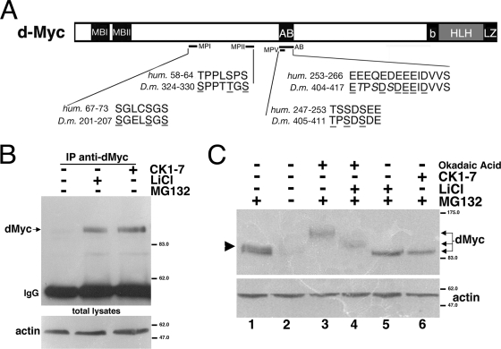
FIG. 1.
Inhibition of GSK3β and CK1 kinases increases dMyc protein level in S2 cells. (A) Schematic representation of dMyc protein with the amino acid sequences of dMyc phosphorylation mutants and homologous sequences in c-Myc. The amino acids changed by site-directed mutagenesis are underlined. (B) Treatment of S2 cells with LiCl and CK1-7 kinase inhibitors stabilizes endogenous dMyc protein. Cells were treated with the indicated inhibitors for 4 h; dMyc was immunoprecipitated from the cell extracts by using anti-dMyc antiserum, and its expression level was analyzed by Western blotting with anti-dMyc antiserum. The position of immunoglobulins is marked on the left. Total lysates were blotted with antiactin for control loading. (C) S2 cells were treated with MG132 (proteasome inhibitor), OA (an inhibitor of PP2A), LiCl (an inhibitor of GSK3β), and CK1-7 (an inhibitor of CK1s). Endogenous dMyc protein was analyzed in total lysates by Western blotting with anti-dMyc antibodies; actin was used as a loading control. The arrowhead on the left represents a dMyc doublet visible in cells treated with MG132. The arrows on the right point to dMyc forms of different electrophoretic mobilities. Molecular mass markers are shown on the right.

