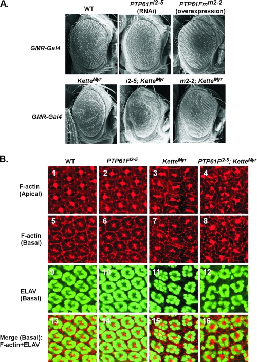
FIG. 2.
The actin cytoskeleton in eye imaginal discs is regulated by Kette and PTP61F. All transgenic constructs were driven by GMR-GAL4 and maintained at 25°C. (A) The phenotype of adult compound eyes was examined by SEM. Ectopic expression of KetteMyr resulted in a rough-eye phenotype. The roughness was enhanced upon RNAi-mediated knockdown of endogenous PTP61F in combination with KetteMyr (i2-5; KetteMyr). In contrast, forced expression of PTP61Fm rescued the KetteMyr-induced eye defect (m2-2; KetteMyr). (B) All images were prepared from the 40-h pupal eye imaginal discs stained with rhodamine-phalloidin (top row and second row from top) and anti-ELAV antibody (third row from top). F-actin organization is shown at both apical (top row) and basal (second row from top) levels, whereas the neuronal pattern of photoreceptor cells is shown at the basal level (third row from top). Merged images of F-actin and photoreceptor cells at the basal level are shown in the bottom row. The staining of F-actin (images 1 and 5) and photoreceptor cells (images 9 and 13) in the WT pupal eye discs reveals the organized pattern of ommatidia. This pattern became disorganized in response to ectopic expression of KetteMyr (images 3, 7, 11, and 15). In GMR-Gal4-driven i2-5; KetteMyr flies, severe defects in the F-actin organization (images 4, 8, and 16) and disturbed localization of photoreceptor cells (images 12 and 16) were observed.

