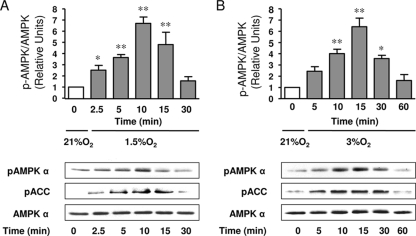
FIG. 1.
Hypoxia activates AMPK in ATII cells. (A) ATII cells were exposed to 21% O2 (white bar) or 1.5% O2 (gray bars) for 2.5 to 30 min, and the levels of AMPK phosphorylated at Thr172 (pAMPK α) and ACC phosphorylated at Ser79 (pACC), as well as the total amount of AMPK α, were measured by Western blot analysis. The graph represents the pAMPK/AMPK ratios. Representative Western blots analyzing pAMPK α, pACC, and total AMPK α are shown. (B) ATII cells were exposed to 21% O2 (white bar) or 3% O2 (gray bars) for 5 to 60 min, and the levels of pAMPK α and pACC and the total amount of AMPK α were determined as described above. The graph represents the pAMPK/AMPK ratios. Representative Western blots analyzing pAMPK α, pACC, and total AMPK α are shown. Values are expressed as means ± SEM (n = 4). *, P < 0.05; **, P < 0.01.

