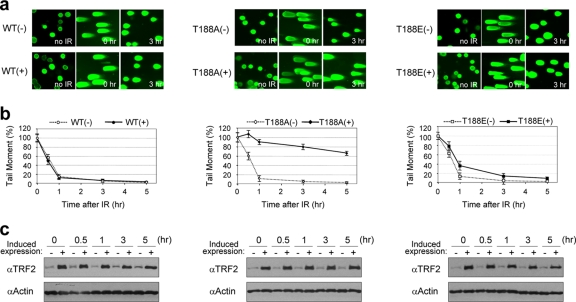
FIG. 3.
Phosphorylation of TRF2 at T188 is critical for the fast pathway of DNA DSB repair. Results for cells without induction (−) and with induction (+) of exogenous TRF2 constructs are shown. WT, TRF2WT construct; T188A, TRF2T188A; T188E, TRF2T188E. (a) Representative neutral comet images. Individual cells are shown with no irradiation (IR), just after irradiation (time zero), or 3 h after 20 Gy X-ray exposure. (b) Neutral comet results shown graphically. The graph shows the percentages of tail moment in TRF2WT, TRF2T188A, and TRF2T188E cell lines either without (−) or with (+) overexpression at indicated times after 20 Gy of X irradiation. Comet images were captured by fluorescence microscopy, and the tail moment was analyzed in at least 100 randomly chosen cells by the TriTekCometScore freeware program. The data represent means ± SD. (c) Expression levels of exogenous, inducible TRF2 proteins. Both endogenous and exogenous TRF2 was detected by anti-TRF2 antibody (4A794) from induced (+) and noninduced (−) cell extracts. Antiactin antibody was used as a gel loading control. α, anti.

