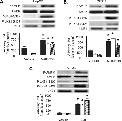
FIG. 3.
Both LKB1 S307 and LKB1 S428 are phosphorylated in metformin- or IBOP-treated cell lines. Confluent cultures of HepG2 (A) or C2C12 (B) cells were exposed to metformin, and vascular smooth muscle cells (VSMC) (C) were exposed to IBOP (1 μM, 60 min). Levels of total and phosphorylated (T172) AMPK, total and phosphorylated (S307) LKB1, as well as phosphorylated (S428) LKB1 were determined by Western blotting. Black bars, P-AMPK; white bars, P-LKB1 S307; and striped bars, P-LKB1 S428. All blots are representative of at least four to six blots from four to six independent experiments. ♣, P < 0.05 versus vehicle.

