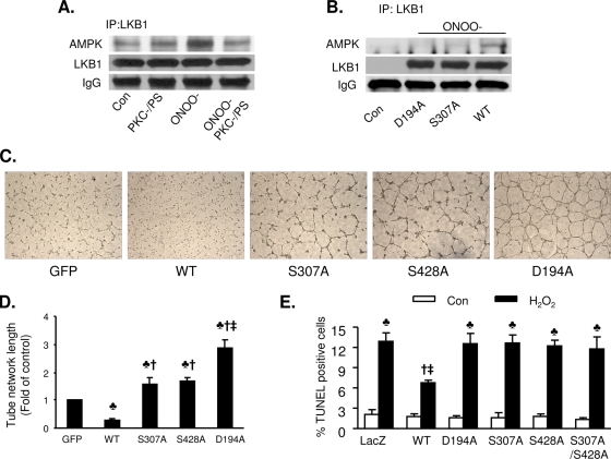
FIG. 8.
Mutation of LKB1 S307 to alanine potentiates angiognesis and increases H2O2-induced apoptosis. (A) Inhibition of PKC-ζ abolishes ONOO−-enhanced coimmunoprecipitation (IP) of AMPK and LKB1. Confluent BAECs were exposed to ONOO− (50 μM) for 15 min. After the treatment, LKB1 was immunoprecipitated using a specific antibody. AMPK and LKB1 were detected by Western blotting. The blot is a representative of three blots from three independent experiments. IgG, immunoglubulin G; Con, control. (B) Mutation of S307 to alanine abolished ONOO−-enhanced coimmunoprecipitation of LKB1 and AMPK. After transfection with WT LKB1 or an LKB1 D194A or LKB1 S307A mutant, A549 cells were exposed to ONOO− (50 μM) for 15 min and LKB1 was immunoprecipitated using a specific antibody. AMPK and LKB1 were detected by Western blotting. The blot is representative of three blots from three independent experiments. (C) Representative images displaying tube formation in HUVECs transfected with adenoviral vectors expressing WT LKB1 or LKB1 mutants at a multiplicity of infection of 25 PFU/cell. (D) Quantitative analysis of tube lengths. n = 5. ♣, P < 0.01 versus GFP; †, P < 0.01 versus WT; ‡, P < 0.05 for LKB1 D194A versus LKB1 S307A or LKB1 S428A. (E) Mutation of LKB1 S307 to alanine abolishes the antiapoptotic effects of LKB1 in response to H2O2. After transfection with WT LKB1 or an LKB1 D194A, LKB1 Ser428A, LKB1 S307A, or LKB1 S307A S428A mutant, A549 cells were exposed to H2O2 (100 μM) for 12 h. Apoptosis was detected by TUNEL staining. n = 6. ♣, P < 0.05 for H2O2 treatment versus the respective control; †, P < 0.05 for WT versus WT plus H2O2; ‡, P < 0.05 for WT plus H2O2 versus H2O2-treated LKB1 S307A or H2O2-treated LKB1 D194A.

