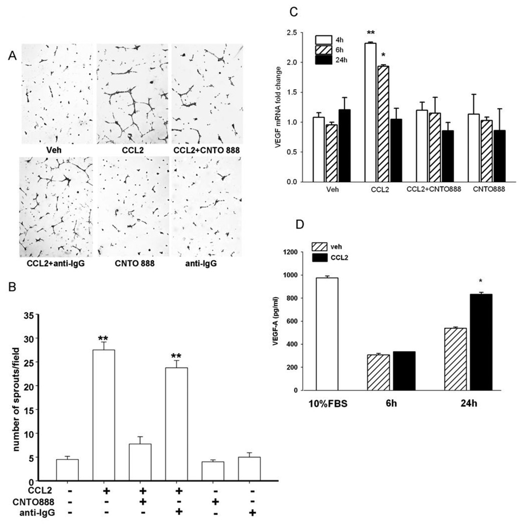
Figure 5. CCL2 has pro-angiogenic effects on endothelial cells through elevated tumor-derived VEGF-A.
(A) Representative images of in vitro vascular sprouting assay of HBMEs in the presence of conditioned media from serum starved PC-3 cells treated with vehicle, CCL2 (50 ng/ml), anti-human CCL2 antibody (CNTO 888) (30 µg/ml) before CCL2, control antibody (anti-IgG) (30 µg/ml) before CCL2, CCL2 antibody and control antibody alone; (B) Conditioned media from CCL2-treated PC-3 cells increased sprout formation in HBMEs while pre-treating PC-3 cells with CNTO 888 abolished the effect; (C) Quantitative-RT-PCR for VEGF-A mRNA expression in serum starved PC-3 cells after 4h (open bars), 6h (hatched bars) and 24h (solid bars) incubation with or without CCL2 and its antibody. Data were standardized to GAPDH. (D) VEGF-A expression in 24h-serum-starved PC-3 cells after 6h and 24h incubation with vehicle (hatched bars) or CCL2 (solid bars). The open bar indicates the level of VEGF-A of PC-3 cells cultured with 10% FBS media.* p<0.01; ** p<0.001 vs. vehicle (Veh); values are mean±SEM; n=3.

