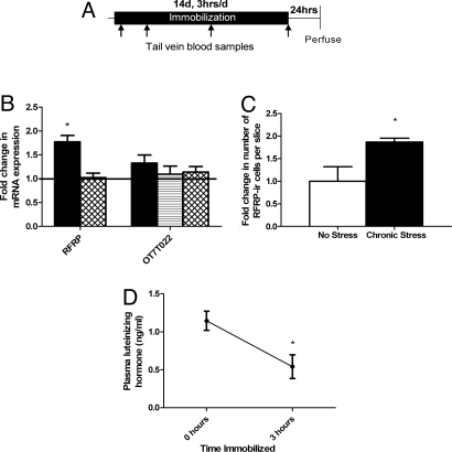
Fig. 2.
Chronic stress effects on RFRP expression. (A) Experimental time line. (B) Gene expression changes in the hypothalamus (solid bars), pituitary (lined bar), and testes (cross-hatched bars) after chronic immobilization, normalized to no-stress control levels. Immobilization led to an increase in hypothalamic RFRP mRNA expression (mean ± SEM, P = 0.007). No change was seen in hypothalamic, pituitary, or testicular RFRP receptor (OT7T022) or testicular RFRP expression (all P > 0.10). (C) Chronic immobilization led to an increase in hypothalamic RFRP-ir cell number in the DMH (mean ± SEM, P = 0.041). (D) Immobilization decreased luteinizing hormone (LH) on the last day of the stressor. Plasma LH levels after stress were significantly lower than those at baseline on day 14 (P = 0.023). *P < 0.05 no stress versus chronic stress or effect of time immobilized within day, as appropriate. †P < 0.10 effect of time immobilized within day. Scale bar, 40 μM.

