
Figure 1.
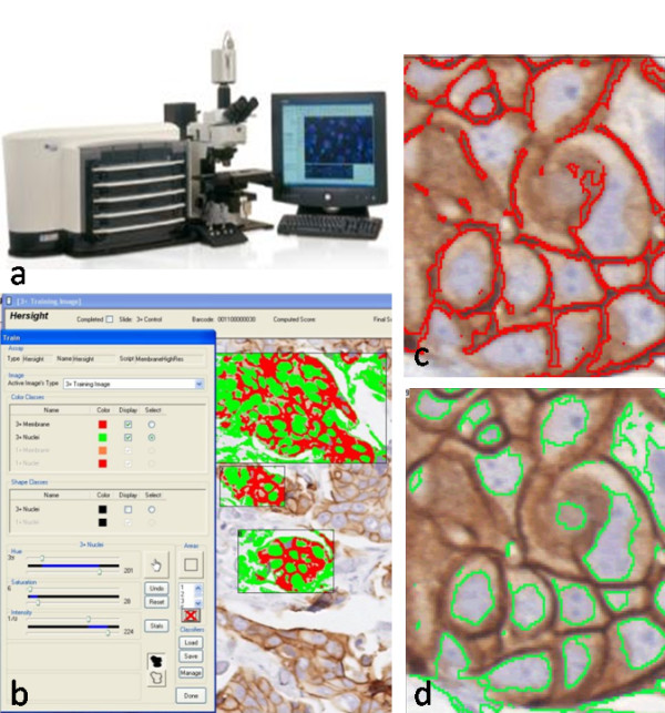
Schematic illustration of automated HER2 scoring. a) Image analysis system Ariol (Applied Imaging Inc., San-Jose, CA). b) Training window displaying the 3+ membrane and nuclear colors with fill mask. c) Outline of membrane as detected by the color classifier for the 3+ membrane color class. d) The border mask of nuclei as detected by the color classifier for the 3+ nuclei color class.