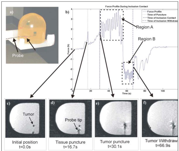
Fig. 9.
(a) Photograph of breast phantom after tumor insertion. (b) Force profile during insertion and withdrawal into phantom breast tissue with tumor. Changing force profile coincides with images from the dynamic scan, depicting (c) initial position (t = 0.0s), (d) tissue puncture (t = 16.7s), (e) tumor puncture (t = 30.1s), and (f) withdrawal from tumor (t = 66.9s). © 2008 IEEE.

