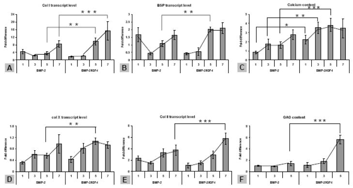
Figure 4.
hMSC osteochondral differentiation in growth factor-gradient silk scaffolds. For all three groups of scaffolds (rhBMP-2, IGF-I, rhBMP-2/IGF-I), scaffolds were sectioned into 7 segments along the direction of growth factor gradient. The results from segment 1, 3, 5, 7 of rhBMP-2 and rhBMP-2/rhIGF-I-incorporated scaffolds are presented. For the scaffolds containing two growth factors, rhBMP-2 concentration increased from segment 1 to 7, while the rhIGF-I concentration decreased. A, B, Bone makers, collagen type I (Col I) and bone sialoprotein (BSP), respectively. C, calcium deposition as weight percentage per (wet) scaffold segment. D, hypertrophic chondrocyte maker, collagen type X (Col X). E, Chondrocyte maker, collagen type II (Col II). F, formation of cartilage specific extracellular matrix material GAG in a scaffold as weight percentage. *Significant differences between the groups (P<0.05). **Very significant differences between the groups (P<0.01). ***Extremely significant differences between groups (P<0.001). Data are Ave.±SD (n = 3–4).

