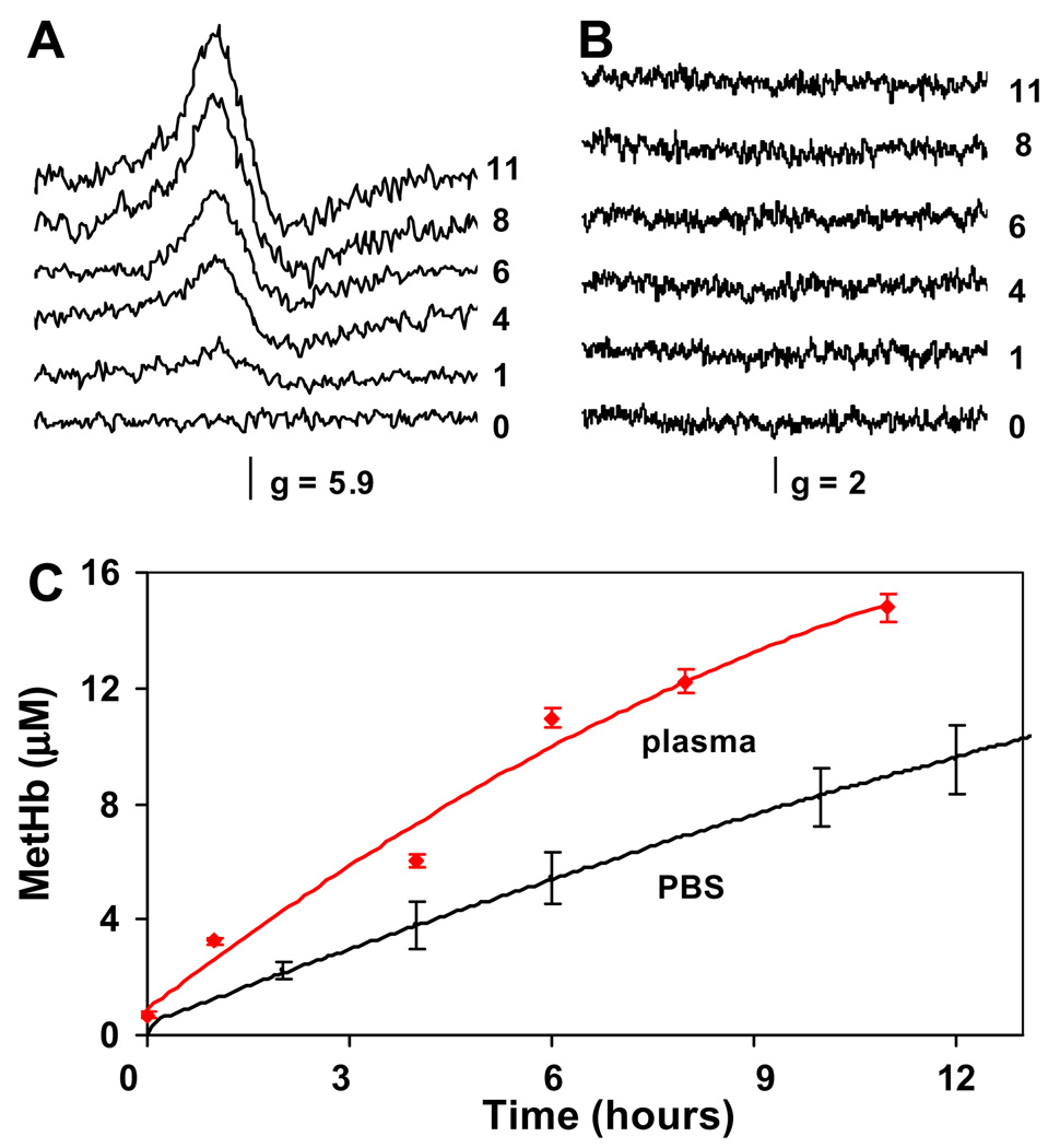
Fig. 4.
(A) MetHb EPR spectra of aliquots taken from the reaction mixture of equimolar ratio of nitrite/oxyHb 1/1 in plasma at different time points indicated by numbers on the right (time given in hours). (B): EPR spectra of the same aliquots as in (B) scanned for the presence of free radical species in g = 2 region. (C) MetHb formation in plasma with added cell-free oxyhemoglobin after nitrite addition as determined using EPR spectroscopy (red line) compared with metHb formation in the same reaction carried on in PBS (pH 7.4) and followed using absorption spectroscopy (black line). Reaction conditions in both cases: oxyHb 30 µM, nitrite 30 µM, 37 °C. (For interpretation of color mentioned in this figure the reader is referred to the web version of the article.)

