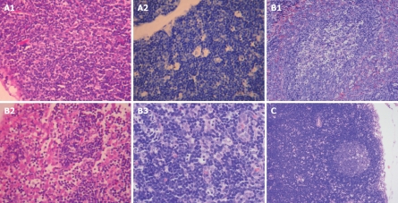
Figure 2.
Pathological changes in lymph nodes of sham-operating group (A), model control group (B), and treatment group (C). A1: 28 d. Mainly normal lymph nodes (HE, × 200); A2: 28 d. No apoptotic cells in lymph node (TUNEL, × 200); B1: 21 d. Focal necrosis in lymphoid follicles and formation of germinal centers (HE, × 200); B2: 21 d. Expansion of lymph sinus, sinus cell hyperplasia and inflammatory cell infiltration (HE, × 200); B3: 28 d. Enlargement and spotty necrosis in germinal centers of lymph nodes, expansion of lymph sinus, hyperplasia of sinus cells and infiltration of neutrophils in lymph sinus (HE, × 200); C: 28 d. Clear follicular structure and fewer necrotic spots in lymph nodes (HE, × 100).

