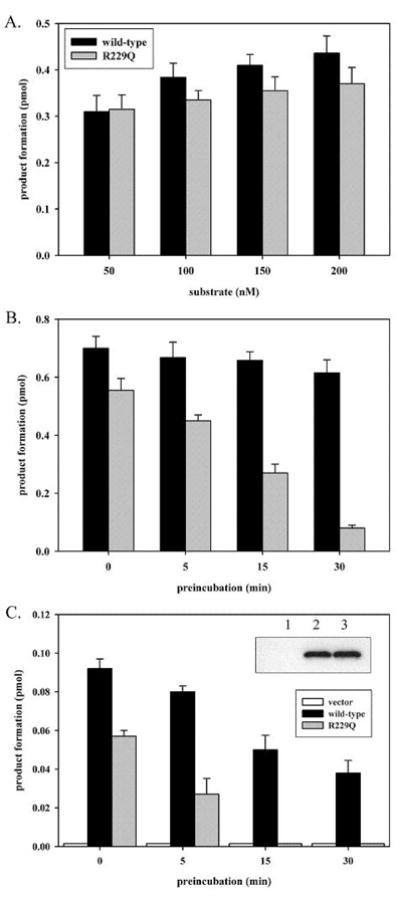
Figure 2.

in vitro and in vivo thermolability of polymorphic R229Q OGG1. (A) Purified wild-type and R229Q OGG1 (12.5 nM) were reacted with 50–200 nM duplex 8-oxoG:C substrate for 5 min. Reactions were performed in 20 mM Tris-HCl, pH 7.4, 100 mM NaCl, and 0.15 μg/μl BSA. (B) Wild-type and R229Q OGG1 at a concentration of 5 ng/μl were preincubated at 37°C for 0, 5, 15 or 30 min. in 20 mM Tris-HCl, pH 7.4, 100 mM NaCl, 0.15 μg/μl BSA prior to being reacted at 12.5 nM with 250 nM 8-oxoG:C substrate for 10 min. (C) KG-1 cells were transfected with pCMV-2B vector or pCMV plasmids encoding N-terminally FLAG-tagged wild-type or R229Q OGG1. Nuclear extracts (2 μg) from KG-1 cells expressing wild-type or R229Q OGG1 at identical levels were assayed for 8-oxoG excision activity with 25 nM substrate for 5 min, with or without preincubating the extracts at 30°C for the indicated times prior to analysis. Panel C inset, anti-FLAG western blot of 2 μg of nuclear extract from KG-1 cells transfected with pCMV vector (lane 1), wild-type (lane 2), or R229Q (lane 3) expressing plasmids. Reactions were terminated and analyzed as described in Materials and Methods. All experiments were performed in triplicate and are shown with standard deviation.
