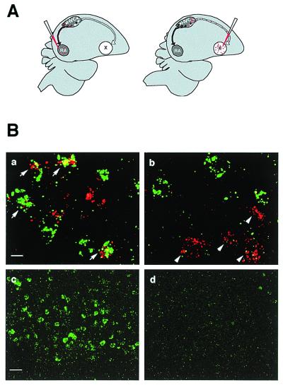
Figure 4.
BDNF mRNA is induced primarily in the RA-projecting neurons in HVC. (A) RA- and Area X-projecting neurons were retrogradely labeled with injections of rhodamine beads into the RA or Area X of each bird (n = 4). (B) The rhodamine signal (red) and the BDNF signal (green) were visualized with confocal microscope at 2-μm photosectioning. (a) Arrows point to neurons backfilled from RA that showed BDNF expression. (b) Arrows point to neurons backfilled from Area X that did not express BDNF. Lower-magnification view of HVC showing the level of BDNF expression was much higher in the singing birds (c) than in the nonsinging birds (d). (Scale bars: 10 μm for a and b; 50 μm for c and d.) See Table 1 for quantification.

