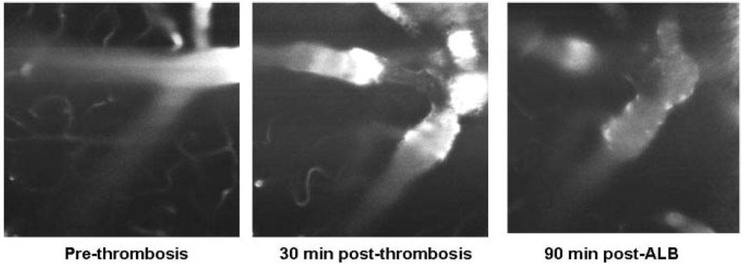
Figure 2.
Left panel: arteriolar field at baseline. Middle panel: at 30 minutes after induction of thrombosis, the thrombosed arteriolar segment is evident by an absence of FITC fluorescence, and focal narrowing is present. Right panel: 90 minutes after ALB treatment, the vasoconstriction is diminished, and the plasma column has become more regular in diameter.

