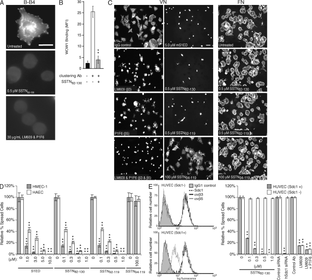
Figure 4.
Inhibition of αvβ3 and αvβ5 integrin activation in HMEC-1 vascular endothelial cells by SSTN. (A) Activation of αvβ3 integrin is blocked by SSTN. HMEC-1s plated for 2 h on hSdc1-specific antibody B-B4 in the presence or absence of 0.5 µM SSTN82-130 or 30 µg/ml of mAbs LM609 and P1F6. Integrin activation is shown by integrin-dependent cell spreading and by staining with the monovalent ligand-mimetic Fab antibody WOW-1. Results are representative of triplicate wells and two independent experiments. Bar, 20 µm. (B) Integrin activation by Sdc1 clustering. Sdc1+ HUVECs in suspension were treated with mAb B-A38 to engage Sdc1 and was treated with or without secondary antibody to cluster the syndecan in the presence or absence of 1 µM SSTN82-130. Quantification of WOW-1 binding via flow cytometry was used to measure the levels of activated αvβ3 integrin. Results are representative of duplicate samples and at least two independent experiments. MFI, mean fluorescent intensity. (C) HMEC-1 spreading on VN is dependent on integrins αvβ3 and αvβ5 and is blocked by SSTN. HMEC-1s plated on VN for 2 h were treated with 30 µg/ml of mAb LM609 (αvβ3 blocker), 30 µg/ml of mAb P1F6 (αvβ5 blocker), or both antibodies, or 5 µM mS1ED, or 0.5 µM SSTN82-130, SSTN92-119, or SSTN94-119 as competitive inhibitors. The SSTN inhibitors were also tested on cells plated on FN as a negative control (right column). Results are representative of triplicate wells and at least two independent experiments. Bar, 50 µm. (D) Quantification of cell spreading. The percentage of spread HMEC-1s and HAECs was quantified after plating for 2 h on VN in the presence of mS1ED or SSTN peptides. **, P < 0.01. (E) Sdc1-negative HUVECs have activated αvβ3 and αvβ5 integrin but are insensitive to inhibitory SSTN peptide treatment. Expression of Sdc1, αvβ3, and αvβ5 are quantified by flow cytometry (left) in two isolates of HUVECs, one of which is Sdc1 negative (Sdc1−). The Sdc1+ and Sdc1− HUVECs are compared in cell spreading assays on VN, using competition with SSTN82-130, silencing Sdc1 expression with siRNA, and blocking αvβ3 alone or both αvβ3 and αvβ5 integrins with blocking antibodies LM609 and P1F6 (right). Results are representative of triplicate wells and at least two independent experiments. Data are presented as means ± SEM. **, P < 0.01.

