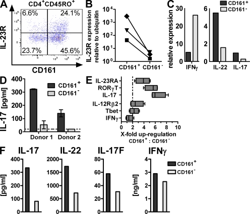
Figure 1.
CD161 expression identifies tissue-resident and circulating Th17 cells. (A–D) LPMCs were isolated from non-CD colon specimens. (A) Flow cytometric analysis for IL-23R expression gated on CD45RO+CD4+ T cells. The plot is representative of five independent donors. (B–D) CD4+CD45RO+ T memory cells were FACS sorted for CD161 expression. il23r (B; data are from three individual donors) or il17, il22, and ifng (C; data are representative of two donors) expression was assessed by qRT-PCR and normalized to ubiquitin. Cells were cultured for 3 d with anti-CD2/anti-CD3/anti-CD28 activation beads for measurement of IL-17 production (D; data are from two different donors). The dashed line indicates the lower limit of detection. (E and F) PBMCs from healthy donors were FACS sorted for CD161+ and CD161− CD4+CD25−CD45RA− T memory cells, and gene expression was profiled (E; data are from four different donors; the dashed line indicates twofold change). The boxes indicate interquartile range with median, and whiskers define minimum to maximum values. Cytokine production was determined after 3 d of culture with anti-CD2/anti-CD3/anti-CD28 activation beads. (F; data are representative of at least three donors from independent experiments).

