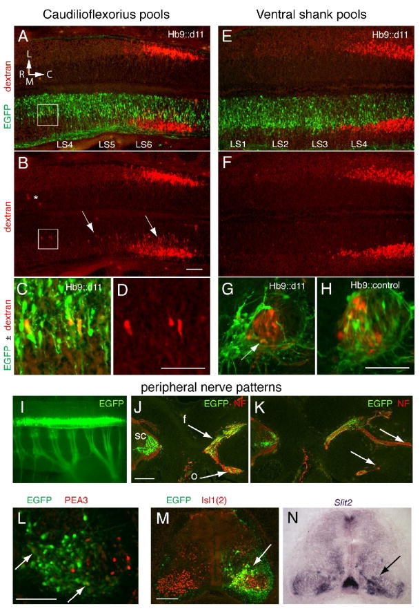
Figure 6.
MNs in rostral LS segments appear to demonstrate a caudal (caudilioflexorius) identity after transfection with Hb9∷d11 but also show abnormalities in cell positioning and axon outgrowth. A. Horizontal section showing caudilioflexorius pools (dextran+ cells) on non-transfected (top) and transfected (EGFP+) sides of the ventral LS cord from a stage 30 Hb9∷d11 embryo. L, lateral, M, medial, R, rostral, C, caudal. B. Dextran alone. On the non-transfected side, few if any dextran+ cells are located outside a major cluster in LS5-6. (The asterisk indicates fluorescence likely to be artifactual as it is diffuse and located well outside the motor column region.) On the transfected side, numerous dextran+ cells are positioned more medially and rostrally than normal (box and arrows). C and D. Boxed area in A and B. On the transfected side, rostrally positioned MNs are EGFP+ and dextran+. E. and F. Ventral shank pools from an Hb9∷d11 embryo. Staining and orientation as in A and B. Dextran+ cells are not present in segments rostral to the main body of the ventral shank pool. G. and H. Transverse sections through motor columns of an Hb9∷d11 (G) and an Hb9∷control embryo (H). In the Hb9∷d11 embryo, most EGFP+ MNs occupy an extreme medial position and only a small number of EGFP+, dextran+ MNs (arrow) are evident. In the Hb9∷control embryo, EGFP+ MNs are more widely distributed in the motor columns, and EGFP+, dextran+ cells, more numerous. I. EGFP expression in cord and limb nerves in whole mount of a stage 27 Hb9∷d11 embryo. J-K. Transverse sections showing femoral (f) and obturator (o) nerve trunk bifurcation (J) and distal branching (K) in an Hb9∷d11 embryo. Arrows indicate the presence of EGFP+ axons at proximal levels in both nerve trunks (J) but their relative absence at more distal levels (K). L. Section through a caudal LS motor column showing extensive Hoxd11 transfection but only a very small number of EGFP+, Pea3+ cells (arrows). M-N. Adjacent sections stained for EGFP, Isl1(2) and Slit2. Slit2 expression is high in medial motor regions where Hoxd11-transfected cells appear to be most numerous (arrows). Scale bars=100μm, except in J-K, 200μm.

